Our interest in this painting goes back many years to when it was mentioned in articles on pioneer surgeons (1 – 3). As we are interested in medical and surgical history, we have provided one another with literature about this painting, for which possible errors in Rembrandt’s anatomical lines has been an object of debate among surgeons and anatomists since the 1920s (4 – 12). The painting was by then world famous after an American cigar merchant had used it to decorate his cigar boxes (Dutch Masters) (3). It was also used on the title page in several editions of the famous Grant’s anatomy and on the cover of the Norwegian Association for Hand Surgery’s anniversary book (13). The Nordic Orthopaedic Association has used it on their folder and homepage for this summer’s congress in Amsterdam.
In this article, we want to concentrate on the surgical anatomical details in the painting in addition to a general discussion of its history.
Rembrandt
Rembrandt Harmenszoon van Rijn was born on 15 July 1606 in the Dutch town of Leiden south of Amsterdam. Leiden lies at the mouth of «the old Rhine» – whence the name van Rijn. Rembrandt was the ninth child in the family, and already demonstrated a painting talent when he went to Latin School in his hometown. He began to study philosophy at the famous University in Leiden, but broke off his studies as early as 1620 and began as an apprentice to the renowned artist Jacob van Swanenburgh. In 1623, he moved to the studio of the master painter of history Pieter Lastman in Amsterdam. Lastman had been in Italy where he had met Caravaggio (1573 – 1610) (14, 15). After six months, Rembrandt finished his apprenticeship in Amsterdam and set up his own studio in his hometown. In 1631, he returned to Amsterdam and settled down for good. There he came in contact with the influential Surgeons’ Guild, and in 1632 he painted his first group portrait Dr. Tulp’s anatomy lesson (fig 1). When the painting was finished, Rembrandt became famous within a few months and, in the following years, received a stream of commissions from citizens and from the court at The Hague (14, 15).
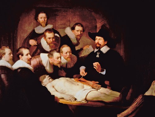
Figure 1 Dr Nicolaes Tulp’s anatomy lesson (1632): Rembrandt’s first group portrait of the surgeons’ guild in Amsterdam or, more precisely, the members of the guild who wanted to pay to be in the painting. It was commissioned by praelector Dr Tulp who thereby got the best position. Photograph © SuperStock/GV-Press
The painting
In this painting, Rembrandt succeeded in producing a lifelikeness that had not previously been seen in this type of group portrait (16). The depicted anatomy lesson is not a superficial motif for portraying members of a guild. On the contrary, with the help of the onlookers’ intense concentration, he places the lesson in the foreground. The picture is strikingly innovative compared with earlier paintings of anatomy lectures, which all had a clear symmetrical construction and where the portrayal of the surgeons was the most important aspect (fig 2). In Rembrandt’s composition, the seven spectators are placed in the left half of the picture: only the lecturer Dr Tulp stands on the right. The corpse is placed diagonally in the painting. All the onlookers, but one, are engrossed in the actual dissection and do not stare at the painter – unlike the norm for this type of painting. Rembrandt bathes the surgeons in a remarkably strong light that comes in from the left. In this way, he makes them all stand out from the surrounding darkness as a unit – so as to be seen as a closed group (7, 14).

Figure 2 Dr. Sebastiaan Egbertsz de Vrij’s anatomy lesson from 1603, painted by Aert Pietersz. Photograph © Amsterdam’s Historisch Museum. Reproduced with permission
In the background to the right, Hartman Hartmanszoon holds a piece of paper where the names of the seven observers are written. Only two of these (Slabberaan and de Wit) were at that time masters of surgery and members of the Surgeons’ Guild in Amsterdam. The others were lesser-known surgeons who had been brought in to share the costs of the painting. Behind stands Frans van Loenen, who looks out at us and appears to direct our attention as viewers to the actual dissection. To the left stands Adriaan Slabberaan, who casts a glance at the open textbook on the right side of the painting. One gets the distinct feeling that he is about to look back at the prepared cadaver after having checked how this is described in the book. Students of anatomy will recognize this habit. The book may well be the famous De Humani Corporis Fabrica by Andreas Vesalius from 1543. Or perhaps the later Anatomia by Adrianus Spiegelius and Julius Casserius from 1627. To the far left is the portrait of Jacob Koolvelt which was added later. He looks straight at Dr Tulp and appears to be listening to what is being said. Some think that someone other than Rembrandt added his portrait later (5, 9).
Matthijs Calkoen, the surgeon nearest Dr Tulp, is staring intensely at Tulp’s left hand. Jacob Block, behind him, is doing likewise, while Jacob de Wit is bending slightly forward to see the dissected corpse that Dr Tulp is lecturing about.
In the picture, a certain discrepancy in size is noticeable between the dissected upper arm in the back, and the intact arm in front (fig 1). Many think that Rembrandt used an already dissected arm as a model (5, 8, 9). He may also have relied on the anatomical drawings of Vesalius or Casserius. X-rays of the painting have shown that the corpse’s left arm ended in an amputated stump. Rembrandt tried to camouflage this by painting a new arm on top (9). Others have thought that Rembrandt did not quite understand what Dr Tulp was actually teaching, and that he later had to correct the picture for this reason (7).
Infrared light has also revealed other changes in the painting. The sheet of paper with the names of the observers was also added later. There used to be an anatomical drawing of forearm muscles and tendons under the list of names. This would have strengthened the dynamics of the painting in that Hartmanszoon had just looked up from the book and completed Slabberaan’s movement in the foreground. The last name on the list, Jacob Koolvelt, is painted in another style than Rembrandt’s, consequently by another hand. It appears that also the person at the top of the painting, Frans van Loenen, was originally painted with a hat that was later erased: probably so he would not to be too dominating in relation to the main character, Dr Tulp (5, 6).
Dr Nicolaes Tulp (1593 – 1674)
Claes Pieterszoon was born in Amsterdam on 9 October 1593 and studied medicine at the University of Leiden from 1611. He practised both as a physician and as a surgeon, and after a while established a successful practice in Amsterdam. During his studies, he changed his name from the very common Claes Pieterszoon to the more scholarly Nicolaus Petreius. In 1621, he established himself in a fashionable area and – influenced by the city’s love of tulips – changed his name to Nicolaes Tulp (tulp = tulip). He decorated his house with a coat-of-arms in the form of a red tulip. Later, when he had become mayor, he stamped hundreds of official documents with his tulip-shaped stamp (17).
Nicolaes Tulp became leader of the city?s Guild of Surgeons in 1628. His inaugural lecture as Praelector Anatomiae was on the relationship between body and soul. In 1636 he published his pharmacopeia and formed the Collegium Medicum. In 1641 he completed his thesis Observatorium medicarum libri tres, cum aenis figuris. The work appeared in a series of revised editions over the next 50 years. His most important anatomical discoveries were his descriptions of the human ileocecal valve (valve ileocecalis) and the lymphatic circulation (vasa lactea). As a surgeon, he was one of the first to describe drainage as a treatment for empyema, and elective trepanation for epidural hematoma. His description of beriberi is regarded as the first in history. In 1650, he led his first official dissection and completed his career as teacher and leader for the city Guild of Surgeons (1, 17).
Dr Tulp was also politically active. As early as 1622, he became alderman and magistrate. He was elected mayor of Amsterdam in 1653 and re-elected altogether three times. He also held a number of other positions and was a member of the city council for more than 50 years (3, 17).
In 1555, the Amsterdam Guild of Surgeons was given permission every year to dissect the body of an executed criminal. The dissections were open to members of the guild, who were actually fined if they did not turn up! They were also open to the general public for a fee. After a while it became a tradition for members of the guild to have their portrait painted at the dissection. A unique collection exists of in all nine such group portraits of surgeons from Amsterdam from the year 1603 to late in the 1700s (5, 16).
In 1632, Dr Tulp was 39 years old and had been a praelector for four years. He wanted to be painted in his «natural surroundings» as his predecessors in the guild had been. An opportunity for the year’s dissection came at the end of December 1631. The 28-year-old criminal Aris’t Kint, also from Leiden, was arrested for armed robbery. His real name was Adriaen Adriaenszoon, but he was called «het Kind» or «Aris’t Kind» (the kid). He had started his criminal career in 1623 when he was arrested for the first time because of theft. He had continued as a criminal and been given a number of sentences. He had finally tried to kill a well-to-do citizen. Because of a previous attempt at murder, he was sentenced to be «hung by the rope». The punishment was carried out on 28 January 1632 and the body was conveyed to the Theatrum Anatomicum. The dissection was performed by praelector Nicolaes Tulp 31 January 1632 with amongst others Rembrandt as observer (3, 17).
What was the topic of Dr Tulp’s lecture?
There are many reasons to believe that the painting shows praelector Tulp giving a lesson on the hand’s muscle function. Tulp is lifting a muscle belly by use of the artery forceps. If you look at the distal end of the tendons, you see the flexor digitorum superficialis (FDS) muscle joined to the middle phalanges of the fingers. The division into the two terminal tendon spindles is clearly seen around the profundus tendons just before the insertion (fig 3).
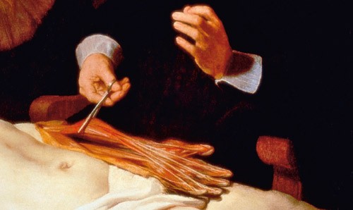
Figure 3 Painting close-up detail with the dissected left upper extremity. Using forceps, Dr Tulp is lifting up the loosened flexor digitorum superficialis muscle. By the fact that the little finger is more flexed than the other fingers (a little more pull in the tendon of the fifth finger than in the other fingers), we understand that the muscle belly (FDS) is being held a little towards the radius. It is this positioning of the muscle belly that has led to the idea that Rembrandt placed the origin of the muscle in the wrong place. Photograph © SuperStock/GV-Press
Rembrandt is said to have misplaced the origin of the muscle at the lateral epicondyle (3 – 12), when it is commonly known to be at the medial epicondyle of the elbow. This has been challenged by others who maintain that Dr Tulp has already loosened the origin in order to demonstrate the effect of the muscle in flexing the finger’s middle joint (PIP joint). This can be seen from certain of the white fibres at the far proximal end, which may indicate that the muscle belly has been loosened from its attachment and Tulp is lifting it up (10). From contemporary anatomy drawings, we can see that this was normal. This is probably also a separate dissected upper arm, which accounts for the somewhat distorted perspective.
In connection with the 400th anniversary of Rembrandt’s birth in 2006, a group of Dutch colleagues dissected an upper arm in order to establish once and for all whether there were anatomical errors in the painting. After this dissection they concluded that Rembrandt really had made anatomical errors (11). But we do not agree with their interpretation. We think Dr Tulp has loosened several of the structures of the forearm (e.g. m. Palmaris longus) in order to free the superficial flexor muscle (m. flexor digitorum superficialis), which he wanted to demonstrate. Rembrandt would surely have made a special effort with his first group portrait and his commissioner, the famous anatomist Dr Tulp, would hardly have allowed him to portray the anatomy wrongly. Several of the other observations made by the Dutch group have also recently been refuted (12).
The fact that Dr Tulp demonstrates the muscle function of the flexor digitorum superficialis can also be seen from the odd way he is holding his left hand. This is no typical gesture of speech, which was often portrayed at the time. His left wrist is fully extended and, with his base joints straight (MCP: metacarpophalangeal joints), he is showing flexion in his middle joints (PIP). He is probably about to pull the muscle belly with his forceps to show the same action on the corpse. In support of this interpretation, we see that the surgeon Matthijs Calkoen, just to the right of Dr Tulp, is also flexing the PIP joints in the same way with his own left hand (fig 1). Rembrandt has also created the illusion of movement in the painting. For whereas Jacob de Wit is bending forward and watching what Dr Tulp is doing with the corpse, Calkoen, to his right, is staring just as intensely at the hand of his master. We know what is about to happen in the next second when Dr Tulp pulls on the tendons. This gives a dynamic climax to Rembrandt’s presentation of functional anatomy, as opposed to the descriptive anatomy common at the time. However, the painting does not show a normal anatomy lecture for the year 1632. This is seen from the fact that the abdomen and chest have not been opened (3, 18). In the 1600s, the perishable inner organs were usually dissected first, and then the extremities. Has Dr Tulp chosen to go directly to the arms? Or have the surgeons positioned themselves in such a way that Rembrandt could draw them before the dissection started and then add in the anatomical details relevant to the lecture later?
In 1543, Andreas Vesalius (1513 – 64) issued (as previously mentioned) history’s most famous anatomical work De Fabrica Humani Corporis – with 700 folio pages and 300 illustrations. The work in seven volumes was printed in Basel and is perhaps the most splendid medical textbook that has ever been published. On the title page, Vesalius is portrayed with a dissected forearm and hand – and, at the same time, holding up a freed m. flexor digitorum superficialis in his right hand (fig 4). The background for Dr Tulp’s dissection may be that he wanted to appear as Vesalius had done a hundred years earlier, and be seen as his successor (3, 5, 6). It is also probable that Rembrandt used Vesalius’ work himself to describe certain details.
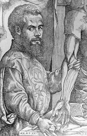
Figure 4 Andreas Vesalius as he was portrayed in a woodcut from 1542. This was used as the title page of his great anatomical work «De Fabrica Humani Corporis». In his right hand he is holding the dissected m. flexor digitorum superficialis. Photograph © Science Photo Library/GV-Press
Conclusion
Rembrandt’s fame and riches continued to grow after 1632, but the death of the prince who had been his most important patron (in 1647) started a long inexorable decline. Even though this phase of his life was also rich in art, his reputation suffered and he went bankrupt in 1656. On 4 October 1669 Rembrandt Harmenszoon van Rijn died in the poor quarter of Amsterdam, aged 63 (14, 15).
