High-pressure glaucoma and normal-pressure glaucoma probably have the same pathogenesis.
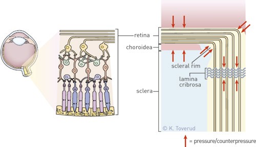
The retinal fibres are compressed during systole, at the same time as the lamina cribrosa with nerve fibres undergo posterior displacement. This traction on the fibres, combined with compression against and over a sharp scleral rim, constitutes an important mechanism.
Normal-pressure glaucoma and primary open-angle glaucoma are virtually identical in clinical terms. A critical analysis of energy and mechanical forces has shown that the papilla with excavated rim in the two conditions has the same causal mechanism (1).
Only the pulse amplitudes at the intraocular pressure peak, Psyst , provide enough energy to cause posterior pulsatile displacement of the lamina cribrosa, while the nerve fibres are compressed in the retina. The nerve fibres are thus subjected to traction over the sharp scleral rim, and at the same time the fibres are subjected to direct compression against the rim. The combination of mechanical forces on the retinal fibres over and against a sharp scleral rim may result in loss of axonal transport and destruction of the nerve fibres. This process is continuous, regardless of whether the intraocular pressure is high or low. Any pathological changes are cumulative.
High-pressure glaucoma and normal-pressure glaucoma therefore probably have the same pathogenesis. This fits well with the view that the load on the connective tissue in the papilla is substantial, even at low intraocular pressures (2).
