A woman in her 60s was referred to a urologist because of recurrent urinary tract infections and haematuria. Assessment revealed an unusual cause of the patient’s complaint.
The diagnostic work-up of macroscopic haematuria with uncertain cause should aim to determine whether the source of the bleeding is the upper or lower urinary tract. Haematuria may be due to benign complaints such as concrements in the upper urinary tract or urinary bladder or malignant complaints such as cancer of the kidney, renal pelvis, ureter, urinary bladder or urethra. Haematuria may also be observed in various kidney diseases and following trauma. The spasmodic back pain described for this patient arouses suspicion of ureter obstruction and possible ureter concrement.
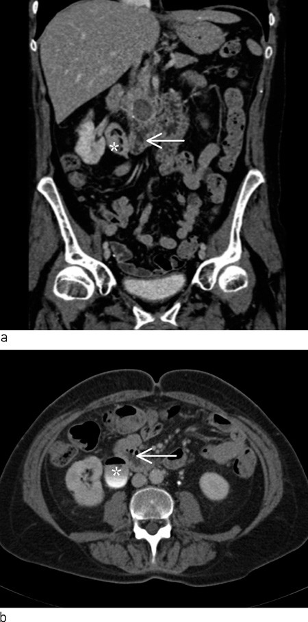
Figure 1 CT urinary tract with intravenous contrast in secretion phase. Coronal (a) and axial (b) reconstruction show close relations between duodenum (arrow) and pronounced renal pelvis with gas and filling defects on right side (star). On the coronal reconstruction, cystic filling is also seen in the pancreas cranially of the duodenum
Pneumaturia (gas in the urinary tract) may be due to a fistula between the bowel or genitals and the urinary tract. This condition may be seen as a complication of bowel cancer, diverticulitis, inflammatory bowel disease or following a surgical procedure in the abdominal cavity (1, 2). Gas may also be found in the urinary tract after instrumentation of the urinary tract or if gas-producing bacteria are present in the urine. In the case of this patient, we had no information regarding episodes of diverticulitis, inflammatory bowel disease or previous surgery that might have made her susceptible to a fistula between bowel or genitals and the urinary tract. The cyst in the head of pancreas, which might be consistent with previous pancreatitis, was initially regarded as a secondary finding.
Her HbA1c value was high, consistent with poorly managed diabetes mellitus. The elevated ALP and GT values were accordingly viewed as non-specific, possibly caused by overconsumption of alcohol.
Up to this point, the work-up had concentrated on detecting a fistula to the urinary tract, but no fistula to the urinary bladder or the upper urinary tract was found. On discharge it was believed that gas-producing bacteria in the urinary tract were a possible cause of the patient’s persistent pneumaturia.
The patient’s pneumaturia disappeared during antibiotic treatment. This strengthened the suspicion that the cause was recurrent infection with gas-producing bacteria.
The patient had persistent pneumaturia, but otherwise little discomfort. The work-up with repeated CT scans had revealed gas in the urinary bladder and collecting system of the right kidney, but had failed to find any cause for her recurrent urinary tract infections or pneumaturia. There were plans for further examination with flexible ureterorenoscopy, which is an endoscopic examination where the ureter and renal pelvis are examined via transurethral access with the aid of a flexible scope. This method makes visual inspection of the ureter, renal pelvis and calyces possible. It is also possible to crush concrements in the urinary tract via laser fibres, extract coagula or concrements or take biopsies by means of instruments that can be introduced through the scope.
Repeated CT scans had not provided a final clarification of the patient’s condition. Gas-producing bacteria were still strongly suspected, and the patient was examined further by means of the planned scan of ureter and renal pelvis with flexible ureterorenoscopy. The patient was admitted for the planned procedure a month later.
Urosepsis is a not uncommon complication of urinary tract procedures, particularly if the procedure takes place during a urinary tract infection. It is therefore advisable as a rule that the patient be completely free of infection prior to instrumentation in the upper urinary tract. In this patient with chronic recurrent urinary tract infections, it would have been difficult to achieve freedom from infection. We therefore decided to operate despite the fact that she had a higher risk of infection. During the procedure, soft structures consisting of protein-rich collagenous tissue and debris consistent with deposits after a chronic urinary tract infection were found in the right kidney.
Discussion
Gas in the collecting system of the urinary tract may be due to instrumentation in the urinary tract, a fistula between the collecting system from the urinary tract and a hollow organ, or infection with gas-producing microbes (3). The most usual cause of pneumaturia is a colovesicular fistula, but it may also be caused by fistulae between other parts of the bowel or genitalia. It may be difficult to detect a fistula, and repeated tests are often necessary. If no fistula is found, infection with gas-producing microbes must be considered as a possible cause. In our patient, chronic urinary tract infection with gas-producing bacteria was the most probable cause of the pneumaturia. The chronic urinary tract infection probably led to the deposition of debris, which in turn helped to maintain the urinary tract infection. The spasmodic back pain experienced by the patient shortly before the initial referral may have been caused by transitory occlusion of the right ureter. In the case of this diabetic patient, we might have considered the possibility of diabetic neuropathy with affection of the bladder, which could have led to increased residual urine and secondary vesicoureteral reflux, which makes patients susceptible to recurrent urinary infections. With this patient, the focus was initially on a haematuria work-up, then on work-up of a possible fistula to the urinary tract. Residual urine was not measured, nor were urodynamic tests or a study of possible vesicoureteral reflux carried out.
Infections in the urinary tract with gas-producing bacteria may be serious, with a high mortality. Infections with gas-producing organisms in the lower and upper urinary tract are called emphysematous cystitis and emphysematous pyelonephritis, respectively. A meta-analysis of 175 patients with emphysematous pyelonephritis revealed a mortality of 25 % (4). The treatment administered previously was often nephrectomy, but with modern treatment modalities, it is possible to achieve the desired effect by relieving the kidney with nephrostomy, antibiotics and good intensive care. Emphysematous pyelonephritis can be classified as type I or type II, depending on whether there are both parenchyma destruction and gas, or whether there is renal or perirenal fluid with gas in the collecting duct system (5). The infections can be divided into four classes, depending on the location of the gas in the CT images (6). Class 1 describes gas in the collecting system, and can be treated with antibiotics. This was the case with our patient. In class 2, there is gas in the renal parenchyma, and in class 3 there is perirenal gas. Class 4 is characterised by bilateral infection or gas in a single kidney. In the more serious infections in classes 2 – 4, the kidney should be relieved by means of nephrostomy or intrarenal drainage. If this is unsuccessful, nephrectomy must be considered.
The most commonly occurring gas-producing bacteria are Escherichia coli and Klebsiella peneumonia (3). In rare cases, Candida infection may also produce gas. Most patients with this problem are in their 60s, and women are affected six times as often as men. Diabetics are over-represented among patients with this problem. In two review articles, 62 – 67 % of the patients with emphysematous cystitis had diabetes mellitus (3, 7). There are several theories concerning the pathogenesis of gas-producing infections. One hypothesis that may explain the high number of diabetics is that a high glucose level in the tissue acts as a favourable substrate, enabling organisms to produce carbon dioxide through natural fermentation (8). Although this cannot explain this condition in non-diabetics, there appears to be general agreement that the combination of potential gas-producing organisms, a high glucose concentration in the tissue and reduced tissue perfusion are associated with the development of emphysematous infections (3, 7). Our patient had both chronic urinary tract infection, poorly managed diabetes and probably also reduced tissue perfusion. Good blood sugar management will probably be conducive to avoiding chronic infection and further episodes of pneumaturia. In conclusion, it may be mentioned that the patient’s pneumaturia, which proved to be an important part of her clinical history, did not emerge in either the referral to a specialist or in her first consultation with a specialist. In cases of recurrent urinary tract infections, it is advisable to ask patients specifically about this symptom.
