A previously healthy woman in her sixties attended the Accident and Emergency department after four days with discomfort and tenderness under her right breast. The skin under the breast tightened whenever she moved her shoulder. Palpation revealed a taut, subcutaneous cord-like induration that extended vertically from the lower part of the right breast to the right side of the abdomen, level with the umbilicus.
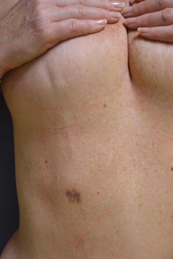
When the Accident and Emergency doctor palpated the breast, the patient felt the cord snap. She subsequently developed a small subcutaneous haematoma on the right side of the abdomen. The cord was still palpable, but no longer taut, and a vertical skin depression had formed caudal to the right mamilla. Ultrasound examination failed to yield clear images of the cord, but certain cross-sections did suggest a 5 mm wide tubular, homogeneous and low-density structure just below the surface of the skin, with echogenicity very similar to that of the surrounding adipose tissue, and absence of a Doppler signal.
A diagnosis of Mondor’s disease was made on the basis of the medical history and clinical findings. Ultrasound examination proved of limited value in this case. The condition was treated conservatively, and clear improvement was seen in both clinical findings and symptoms over the course of a few days.
The photograph shows the subcutaneous induration in the lower part of the right breast and at the level of the umbilicus, a vertical skin depression in the right breast, and the subcutaneous haematoma.
Mondor’s disease was described by the French surgeon Henri Mondor in 1939. It is a sclerosing, superficial thrombophlebitis of the subcutaneous veins on the anterolateral thoracoabdominal wall (1). The condition is rare – approximately 500 cases are described in the literature, mostly in the form of case reports. Possible risk factors are trauma, muscular strain, surgery, breast biopsy (2), venous compression from clothing/bandages and intravenous drug use.
As with our patient, however, in many cases there is no known causal trigger. Treatment is conservative. Resolution can be expected over the course of 1–2 months, and symptoms can be relieved with non-steroidal anti-inflammatory drugs. The connection to breast cancer is unclear, but there should be a low threshold for breast cancer diagnostics in the event of clinical suspicion or known risk factors for breast cancer.
