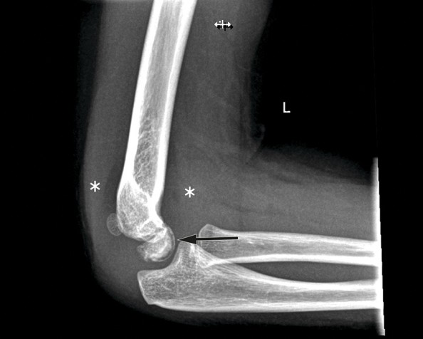
X-ray radiograph showing a radiolucent line (black arrow) corresponding to small gas bubbles visible in the same location on a CT scan (white arrow) – the so-called vacuum phenomenon. Elevation of the anterior and posterior fat pads (marked by asterisks) is a sign of joint effusion. Slight irregularity of the articular contour of the ventral capitulum can also be seen. The findings are typical of Panner’s disease.
This X-ray radiograph of the left elbow shows elevation of the fat pads as a sign of joint effusion (marked with asterisks), but no visible fracture or luxation. A thin strip of reduced subchondral bone density can be seen in the ventral capitulum (black arrow); on the CT scan, this strip corresponds to several small gas bubbles in the same location – the so-called vacuum phenomenon. The articular contour of the ventral capitulum is also irregular. These findings are typical of Panner’s disease.
The images are from a girl of late kindergarten age who was brought to the doctor after four days of pain and reduced mobility in her left elbow in the absence of any known trauma. The patient had had two episodes of radial head subluxation, often referred to as a ‘pulled elbow’, on the same side, the most recent being two years ago. On examination she did not appear to be in pain, but showed a 20–30° reduction in passive and active extension of the elbow joint, and slightly reduced supination. Findings were otherwise normal.
Panner’s disease is an osteochondrosis of the capitellar ossification centre (1). The disease was first described in 1927 by the Danish radiologist Hans Jessen Panner, who observed radiographic changes in the capitulum of a young adult similar to those previously described in cases of osteochondrosis of the hip epiphysis (Calvé-Legg-Perthes disease) (2). The condition is rare and most often occurs in boys under ten years of age. The aetiology has not been fully characterised, but the condition has been associated with trauma to the elbow or repetitive valgus stress (3).
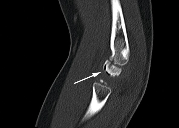
CT scan showing the vacuum phenomenon (white arrow).
Typical clinical findings in Panner’s disease are pain, swelling, stiffness and reduced range of motion in the elbow joint. Morphological changes can be seen on X-ray, along with contour deformity, collapse and increased density of the capitulum (1). The subchondral vacuum phenomenon is a rare finding, but is highly specific to this condition. The vacuum phenomenon, which represents subchondral gas formation, is a sign of bone ischaemia and may indicate aseptic necrosis (1).
The main differential diagnosis of Panner’s disease is osteochondritis dissecans, which typically occurs in older children and young persons aged 10–20 years. It is unclear whether Panner’s disease and osteochondritis dissecans are two different conditions or a continuum of the same condition. Osteochondritis dissecans is more often associated with the formation of intra-articular loose bodies, and with a longer disease course and the more frequent need for surgical intervention (3). Treatment of Panner’s disease is usually conservative with rest and possibly immobilisation, and the condition typically resolves without sequelae.
