Acute appendicitis usually begins with diffuse abdominal pain that migrates to the lower right quadrant and is followed by peritonitis. The clinical picture of the patient presented here also included a painful mass in the right groin.
A woman in her eighties presented to the emergency department with a 1.5-day history of abdominal pain. She had a temperature of 38.4 °C, heart rate of 106 bpm, respiratory rate of 24 breaths/min, and blood pressure of 107/60 mmHg. She had comorbid COPD (GOLD 3), polymyalgia rheumatica and temporal arteritis, and her regular medications included prednisolone 2.5 mg × 1. Ten years earlier, she had undergone open surgical repair of a left inguinal hernia with mesh implantation. She lived at home and coped well with day-to-day activities.
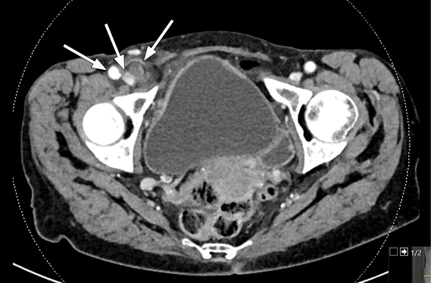
Figure 1 Axial CT scan showing a superficial tubular structure in the right fossa with adjacent adipose tissue remodelling, including thickening of the peritoneum behind. The arrows show (from the left) the femoral artery, femoral vein and appendix.
In the emergency department the woman felt nauseated and unwell, and abdominal examination revealed localised peritonitis in the lower right quadrant. She had a palpable swelling in the right groin of about 1.5 cm in size, which was tender and hard and could not be moved. Blood tests showed CRP (C-reactive protein) 182 mg/L (0–5) and leukocytes 14.9 g/L (3.5–10.0).
On account of suspected appendicitis, CT abdomen with contrast was performed and revealed a superficial tubular structure in the right fossa with adjacent adipose tissue remodelling, including thickening of the peritoneum behind. The scan was interpreted as showing an inflamed appendix; the wall of the appendix was also somewhat poorly defined in places, suggesting perforated appendicitis. In addition, a mass was seen in the right inguinal canal with a small gas bubble. Although there was no definite communication with the appendix, it was nevertheless likely that the appendix had herniated into the femoral canal.
Owing to suspected perforated appendicitis, the woman was given intravenous antibiotics in the form of ampicillin 2 g × 4, gentamicin 320 mg × 1 and metronidazole 1.5 g × 1 prior to laparoscopy.
The distal part of the appendix was incarcerated within the abdominal wall. Because of the tightness of the femoral ring, it was not possible to reposition the appendix laparoscopically, and the operation was therefore converted to an open hernia repair.
The repair was performed without complications, the hernia sac was resected and the peritoneum closed with non-absorbable sutures. The remainder of the procedure was performed via laparoscopy. The distal appendix was found to be gangrenous and to have a small perforation; it was therefore removed, and the area irrigated with copious amounts of sterile saline.
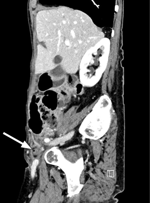
Figure 2 Sagittal CT scan. The arrow shows a mass in the inguinal canal with a small gas bubble.
The patient was hospitalised for five days, during which time her CRP levels decreased from 182 mg/L to 41 mg/L and leukocytes from 14.9 g/L to 10.3 g/L. She was discharged to a short-term placement in a nursing home because of reduced general condition, and because she required assistance with mobilisation. At this point, her antibiotics were changed to oral trimethoprim/sulfonamide 2 tablets × 1 and metronidazole 400 mg × 3 for two days, to give a total of seven days of antibiotic treatment.
Histopathological examination of the appendix confirmed acute transmural inflammation, with no evidence of malignancy.
Discussion
A femoral hernia that contains the vermiform appendix is referred to as de Garengeot’s hernia, after the French surgeon René-Jacques Croissant de Garengeot who first described the condition in 1731 (1). A slightly more common variant of a hernia containing the vermiform appendix is Amyand’s hernia, in which the appendix is found within an inguinal hernia (2).
De Garengeot’s hernia is rare, and only 222 cases have been described worldwide (2). It accounts for 0.5–5 % of all femoral hernias. Acute appendicitis in the hernia sac is even less common, with an estimated prevalence of 0.08–0.13 % (1, 2).
De Garengeot’s hernia occurs mainly in women, and the average age of those affected is around 70 years (1, 2, 4). Patients often present with pain, or a painful lump, in the right fossa or groin, sometimes with overlying erythema. The condition can be difficult to diagnose, and peritonitis caused by underlying appendicitis can be masked because the narrow hernia sac prevents the spread of inflammation to the rest of the abdomen (1, 2, 5).
It is unclear whether the appendix first becomes inflamed and then enters the hernia sac, or whether the appendix enters the hernia sac, becomes strangulated and thereafter inflamed. It has been shown that a large and mobile caecum can push the appendix into the femoral canal.
The condition is often diagnosed during surgery, but can be detected pre-operatively on CT abdomen. CT is the best modality for visualising de Garengeot’s hernia as it can reveal the location and contents of the hernia. The femoral canal is medial to the femoral vein and posterior to the inguinal ligament (1, 4).
De Garengeot’s hernia with appendicitis requires emergency surgery with both appendectomy and hernia repair to prevent fulminant sepsis and complications such as strangulation, perforation and abscess (3, 5). The surgery can be technically challenging and a number of surgical methods have been described, both laparoscopic and open. There are too few case reports to be able to recommend any specific technique, and the choice of method must therefore be tailored to the individual patient and will also depend on the experience of the surgeon (1, 2). Hernia repair with insertion of mesh should be avoided in cases of infection, as the mesh itself can become infected, with the risk of a chronic foreign body reaction (1, 3, 4).
